

3025 Hamaker Court Suite 101
Fairfax, VA 22031

Epiretinal Membrane | ERM | Cellophane Maculopathy
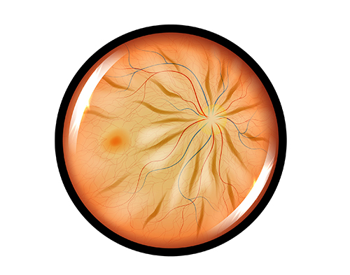
An epiretinal membrane (ERM), also known as a macular pucker or cellophane maculopathy, is a condition characterized by the formation of a thin, transparent layer of fibrous tissue on the surface of the retina at the macula (the central part of the retina responsible for sharp, detailed vision). This membrane can distort and wrinkle the macula, affecting central vision.
The exact cause of epiretinal membranes isn’t always clear, but they often develop due to changes or disruptions in the vitreous gel inside the eye. As the vitreous shrinks or separates from the retina with normal aging, it can cause cells or tissue to be pulled off the retina’s surface, leading to the formation of a membrane.
Other factors contributing to ERM formation may include eye trauma, inflammation, diabetes, or previous eye surgeries.
Many people with epiretinal membranes may not notice any significant changes in their vision. However, in some cases, an ERM can cause certain symptoms.
Symptoms of ERM
Blurred or distorted central vision
Difficulty reading or seeing fine details
Straight lines appearing wavy or bent
Mild or moderate vision loss in the affected eye
Diagnosing an Epiretinal Membrane
Diagnosing an epiretinal membrane typically involves a comprehensive eye examination,
including a dilated eye exam, optical coherence tomography (OCT) to visualize the retina’s layers and detect any membrane presence or changes, and other imaging tests to assess the severity and impact on vision.
Treatment for an Epiretinal Membrane
Treatment for an epiretinal membrane may not be necessary if it doesn’t significantly affect vision or daily activities.
However, if vision impairment becomes bothersome or affects quality of life, surgical intervention may be considered. The primary treatment for an epiretinal membrane is a surgical procedure called vitrectomy combined with membrane peeling.
Vitrectomy for Epiretinal Membrane
During vitrectomy surgery, the vitreous gel is removed, and the fibrous tissue (epiretinal membrane) is delicately peeled off the retinal surface using microsurgical instruments.
After removing the membrane, the surgeon replaces the vitreous gel with a saline solution or gas bubble to help the eye heal and maintain its shape.
Recovery from epiretinal membrane surgery varies from person to person. Some individuals may experience improved vision relatively quickly, while others may take more time to notice visual improvement.
Regular follow-up appointments with your eye doctor are essential to monitor the healing process and vision recovery after surgery.
© 2026 Capital Eye Consultants. All rights Reserved - Accessibility Statement - Privacy Policy - Sitemap
Powered by